Breast PET/MRI Insert Prototype
We aim to use the combination of the complementary detection sensitivities of Positron Emission Tomography (PET) and Magnetic Resonance Imaging (MRI) to improve the diagnostic accuracy in breast cancer. The current whole-body clinical PET/MRI scanners were designed for a wide range of applications, not solely for breast imaging. This causes limitations in spatial resolution and detection sensitivity for imaging of the breast. Our goal is to overcome these limitations by the development of a breast-specific PET insert for a state-of-the-art clinical whole-body PET/MRI scanner. The concept proposed by our group further utilizes coincidences between the breast PET insert and the whole-body PET/MRI scanner. This enables a high precision for imaging of the axillary lymph nodes and metastasis outside of the breast which are essential for the classification of breast cancer and subsequent treatment guidance.
The breast-specific PET insert operates with high spatial resolution due to smaller scintillation crystals (1.51×1.51×10.00 mm³) compared to those in whole-body PET scanners (4.0×4.0×20.0 mm³). This difference allows for finer sampling and higher resolution imaging. The system captures three types of coincident events: internal (within the breast PET insert), external (within the whole-body PET scanner), and mixed (between the breast PET insert and the whole-body PET scanner). Mixed coincidences, which occur in regions like the axilla, leverage the spatial resolution benefits of both systems, providing an effect known as the virtual pinhole effect. We are developing a novel framework that jointly reconstructs these coincidence types, incorporating Depth of Interaction (DOI) and Time of Flight (TOF) information. This innovative approach enhances the diagnostic capabilities and accuracy of breast cancer imaging, aiding in more precise staging and individualized therapy planning.
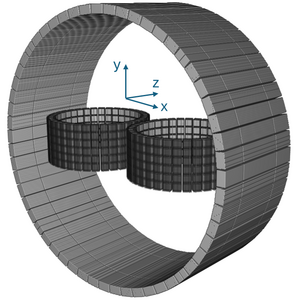
Proposed breast PET insert inside a Whole-body PET scanner.
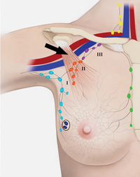
Level I, II, and III axillary lymph nodes of the lymphatic drainage of the breast (by Chang et al, Radiology, 295(3), 500-515, 2020).
Grants
The project is financially supported by the German Research Foundation (DFG) under grant agreement no. 508064995 and NHR Center NHR@ZIB project no. shb00004.

Cooperations
- Eberhard Karls Universität Tübingen
- Universitätsklinikum Tübingen
- Werner Siemens Imaging Center
- Max Planck Institute for Biological Cybernetics